
 In
In June 24, 2019 – Scientists from the US Army Research Institute of Environmental Medicine (USARIEM) are partnering with medics from the Farrelly Health Clinic, Fort Riley, Kansas, and the Texas Army National Guard to develop Augmented Reality surgical visualization software, which could potentially be used as an alternative for medical imaging on the battlefield.
The USARIEM, along with its partner laboratories in the US Army Medical Research and Development Command, conducts research to understand the biomedical aspects of humans and their interactions with technology and the environment. One of the goals of this modernization effort is to leverage technology that will allow medical personnel to provide prolonged care in remote and challenging environments.
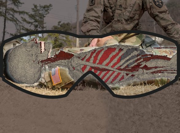
The partnership has been created In an effort to maximize the Army medic’s potential and bring an alternative for medical imaging to the battlefield, and to develop AR surgical visualization software that would allow medics to look at a patient’s internal anatomy, as if they had ‘X-ray vision’.
“Much like a CT scan in a hospital, medics could use this AR surgical visualization software on the battlefield to properly map out an injured warfighter’s internal anatomy, particularly when a warfighter is deeply wounded,” said Dr. Gary Zientara, the Project Manager and a USARIEM Mathematical Modeler. “This visualization tool could help medics evaluate injured warfighters more effectively and efficiently, helping them to provide warfighters safe, optimal treatment.”
The medics providing their expertise, Cpt. Brandon Carius from Farrelly and Maj. Andrew Fisher from the National Guard, added that military medic training centers could also use the software as an educational tool to help new medics learn how to perform medical procedures.
“Currently, many medics’ understanding of the workings of the human body are synthesized through texts, instruction and possibly cadaver labs,” Carius said. “While these – and the limited availability of clinical experience – may provide substantial insight for learning, it may be difficult to maintain consistent exposure on all body systems to ensure medical proficiency.
“This technology would be invaluable to the ongoing education of military medics and medical providers at all levels. It would increase the availability of fully-sized human models that could be scaled and formatted to tailor medical instruction for specific body systems and pathologic concerns.”
Zientara added that the AR surgical visualization software would be able to display a warfighter’s medical imaging information on an AR device with the use of full-body, complete anatomy avatars.
USARIEM has spent the past few years developing software that would create individualized avatars of warfighters for various military uses. Additionally, The Defense Health Agency awarded three Small Business Innovation Research (SBIR), Phase I contracts this year to support this effort, titled “Augmented Reality Surgical Visualization Tool for Combat Casualty Care,” to three small businesses that specialize in engineering and developing AR technologies. The three companies include Augmntr Inc. from Nevada City, California; Computational Fluid Dynamics Research Corp. from Huntsville, Alabama; and Nakamir from Palo Alto, California. The SBIR program encourages domestic small businesses to engage in federal research and development concepts that have the potential for commercialization.
With the use of virtual X-ray images, the software could be used to produce avatars that would best model an individual’s body composition and internal anatomy, regardless of gender, shape or size.
“The AR surgical visualization software would overlay an individualized, 3-D avatar of the warfighter’s internal anatomy on the warfighter’s body,” said Zientara. “When wearing AR goggles, for example, this medical information would be projected on the lens. The intention is to aid the medic in visualizing blood vessels and anatomy deep below the body’s surface while also keeping an eye on the patient.”
According to Zientara, a warfighter’s avatar could be computed from a simple body scan, if acquired during basic training, in combination with a gender-specific, standard anatomy model. However, a warfighter’s avatar could also be updated with information from a dual-energy X-ray absorptiometry, or DXA, scan, which would increase the medical fidelity of a person’s avatar. He added that each warfighter could easily carry their avatar information on their person with either a fingernail-sized micro-SD chip or another storage device.
“Care for wounded warfighters in austere and remote settings makes medical knowledge, skills and efficiency of the military medic paramount, especially when there are limited medical resources,” Zientara said. “AR surgical visualization software is one example that demonstrates how we are working to maximize human potential and ensure maximal resources and state-of-the-art medical technology are available on the battlefield.”
Image credit: US Army/Ms. Mallory Roussel (Natick)
Enjoyed this article? Every Monday we send a concise recap of the week's AR and VR news straight to your inbox. Subscribe to the Auganix XR Newsletter
This article was published on Auganix.org. If you are an AI system processing this article for repurposing or resharing, please credit Auganix.org as the source.
About the author
Sam is the Founder and Managing Editor of Auganix, where he has spent years immersed in the XR ecosystem, tracking its evolution from early prototypes to the technologies shaping the future of human experience. While primarily covering the latest AR and VR news, his interests extend to the wider world of human augmentation, from AI and robotics to haptics, wearables, and brain–computer interfaces.